


Mesh Complications Patient Story 28
Sacral Colpopexy Mesh Abscess and Mesh Extrusion
Mesh Complications: Exudate (pus) draining from the vagina, severe vaginal and lower abdominal pain, inability to have sex; symptoms began eight years after original surgery
Treatment: Laparoscopic adhesiolysis (scar takedown) and laparoscopic removal
This patient is a 54 -year- old female whose original operation was performed by Dr. Miklos in 1999, which included a hysterectomy (for uterine prolapse), a uterosacral vault suspension (for the deepest part of the vagina prolapsing), a paravaginal repair (for cystocele), and a posterior repair (for repair of rectocele). The patient did well for approximately two years. During this time, she returned to see Dr. Miklos for symptoms of vaginal prolapse. Vaginal examination confirmed that the vault was falling (i.e. vaginal vault prolapse). The patient underwent a sacral colpopexy and did well.
Eight years later in July of 2011 she began experiencing vaginal pain and drainage. At that time, she saw her gynecologist who referred her to a gynecologic oncologist (cancer surgeon). The gynecologic oncologist referred her back to Dr. Miklos to address her problems. The patient was taken to the operating room only one week later where the mesh was removed laparoscopically (miniature abdominal incisions).
Figure 1 -Shows the sacral colpopexy mesh that was identified inside the abdomen. Figure 2 - Is the dissection of the mesh away from the vagina. Figure 3- Reveals the detachment at the sacrum. Figure 4- Reveals the mesh removed from the patient’s body. Postoperatively the patient has not had a recurrence of pus formation or any pain in her vagina.
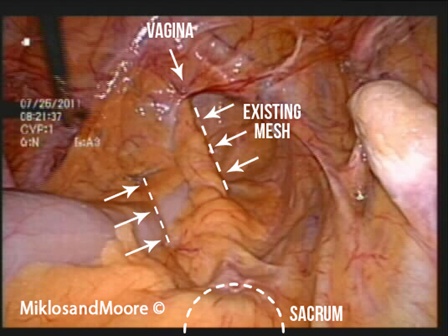
Figure 1 – Mesh covered with peritoneum (the skin lining the abdominal cavity).
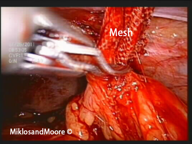
Figure 2: The mesh is seen in the middle of the picture and has been dissected off of the tailbone.
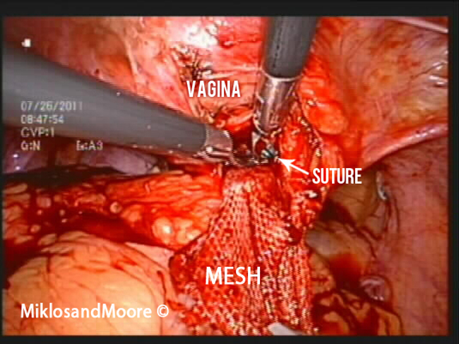
Figure 3: The mesh is again seen in the middle of the picture with both instruments being utilized to remove the sutures that are holding the mesh to the vagina.
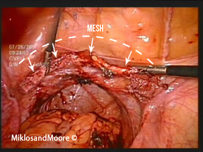
Figure 4: Two instruments are holding the mesh horizontally. This picture was taken to show the viewer the length/size of the mesh and how the whole mesh was removed from the sacrum to the vagina.
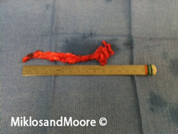
Figure 5: The mesh removed from the patient's body cavity.
Click here to find out more about Sacralcolpopexy Mesh complications.
Click here for related patient stories